References & Reviews from our Customers
Customer References
Chengxi Zhu (Todd),
Cambridge Advanced Imaging Centre,
University of Cambridge Anatomy Building ,
Cambridge UK
"Microscope Heaters 设计的显微镜恒温箱已经应用于剑桥大学多台显微系统上。该恒温箱能快速达到设定温度,恒温效果优异。 Microscope Heaters 的同时提供了优异的售后服务,在恒温箱交付一年内,进行了免费的系统升级,使得性能更佳贴近我校要求。"
"The microscope incubators designed by Microscope Heaters have been used in many microscope systems at the University of Cambridge. The product can reach the set point temperature rapidly whilst maintaining thermal equilibrium within the incubator. Microscope Heaters offers an outstanding after-sales service. It helped to upgrade the microscope incubator for free within the one year after delivery to meet our requirement.""
Giacomo GROPPLERO, PhD,
Macromolecules and Microsystems in Biology and Medicine (MMBM) ,
Institut Curie - Centre de Recherche, UMR 168 ,
Institut Pierre Gilles de Gennes pour la Microfluidique (IPGG) - Paris
"Nous utilisons la chambre d'incubation de Microscope Heaters pour de l'imagerie de cellule de mammifères par microscopie confocal spinning disk……. En plus de l'excellente stabilité en température et de l'absence de vibration, Tqui sont cruciales pour les expériences en molécule unique,……"
Dr. Jens Eriksson,
Manager, Sellin Imaging Platform,
Department of Medical Biochemistry and Microbiology,
Uppsala University, Sweden
"I'm very pleased with our microscope heating system from Microscope Heaters. At our facility we are routinely running 72 h+ long time lapse experiments on primary cells without any issues with it. What really sets the system apart from any other that I have been working with over the past 10 years, is that it is completely silent. As any microscopist knows, the heating system is often the loudest component in a live cell microscopy setup. Because the microscope room is my main working environment, I find it quite pleasant, and probably good for my hearing in the long run, not have the constant noise of a loud convection fan in my ear."
Professor Klaus Suhling,
Kings College London
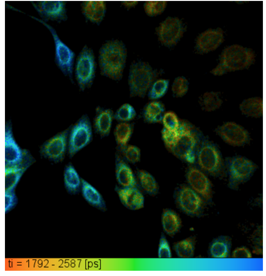
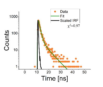
"Fluorescence lifetime image of mitochondria-targeted fluorescent molecular rotors in human epithelial corneal (HCE) cells at 37 degrees C and 5%. [2] The false colour scale represents the fluorescence lifetime from 1.79ns (red) to 2.59ns (blue). A representative fluorescence decay is shown on the right.
We use a Microscope_Heaters Heated Insert and CO2 Controller system to study fluorescent molecular rotor dyes in living cells.[1] These dyes are based on bodipys, and change their fluorescent lifetime as a function of viscosity. Fluorescence lifetime imaging (FLIM) of them allows us to get images of viscosity in cells. We have used fluorescent molecular rotors targeted to mitochondria in human epithelial corneal (HCE) cells at 37 degrees C, which reveals a variability of mitochondria viscosity in livings cells under these conditions.[2]
The Heated Insert and CO2 Controller system from Microscope Heaters is great, as it is small, reliable and portable, so we are easily able to move it across several microscope platforms. Performance and company support in optimizing the system has been excellent. We would highly recommend this system to any researchers using live cell imaging."
Alan Wainman,
Raff Group,
University of Oxford
"Developed for the University of Oxford to image Drosophila embryogenesis, and maintain precise temperature control at 18,22,25 and 30 oC over a 10-24 hour period. Better than 0.2oC accuracy is required and maintained. The system heats the sample, and cools the sample when required."
Testing out our new Heater Cooler System from @cell_viability. Maintaining constant temperature is important to keep our embryos happy! Image credit @caballe_anna pic.twitter.com/US0b3JzQAe
— Raff Lab (@JRafflab) March 22, 2019
Dr Emanuel Derivery,
MRC LMB Cambridge
"Nous utilisons la chambre d'incubation de Microscope Heaters pour de l'imagerie de cellule de mammifères par microscopie confocal spinning disk, ainsi que pour des expériences en lécule unique par microscope à onde évanescente."
"En plus de l'excellente stabilité en température et de l'absence de vibration, Tqui sont cruciales pour les expériences en molécule unique, ce système offre un encombrement minimal sur notre table optique car les éléments chauffants sont petits et que ce système ne repose pas sur des gros tuyaux comme les systèmes concurrents."
Andrew Jefferson,
Micron Imaging Facility,
University of Oxford
"We've been using the Microscope Heaters Objective Heater on our Perkin Elmer Spinning Disc/ Olympus IX-81 microscope with impressive results. Easy to fit and set up, Provides excellent thermostability during extended time course imaging at 37 oC."
Jamie Wheeler,
Foster Group,
University of Oxford
"We study early-stage biofilm formation in the opportunistic pathogen Pseudomonas aeruginosa. This whole process is highly temperature sensitive. Microscope Heaters have designed a microscope incubation chamber highly tailored to our incubation and imaging needs. Crucially, the incubation chamber has dual-capability: it can both heat and cool our samples. It has proved to be extremely reliable and effective, guarding against quite dramatic fluctuations in room temperature."